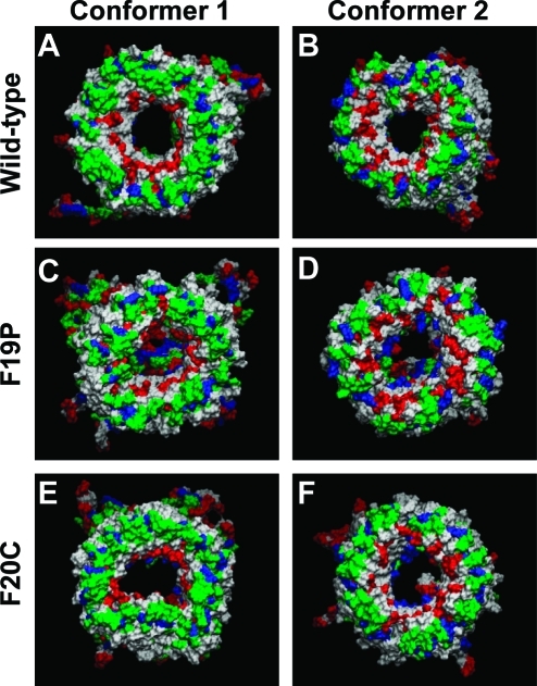
Figure 4.
Cartoons representing snapshots of the averaged Aβ barrel structures over the simulations for the (A) conformer 1 and (B) conformer 2 wild-type Aβ1–42 barrels, (C) conformer 1 and (D) conformer 2 F19P barrels, and (E) conformer 1 and (F) conformer 2 F20C barrels. In the channel structures, hydrophobic residues are colored white, polar and Gly residues are green, positively charged residues blue, and negatively charged residues red.

