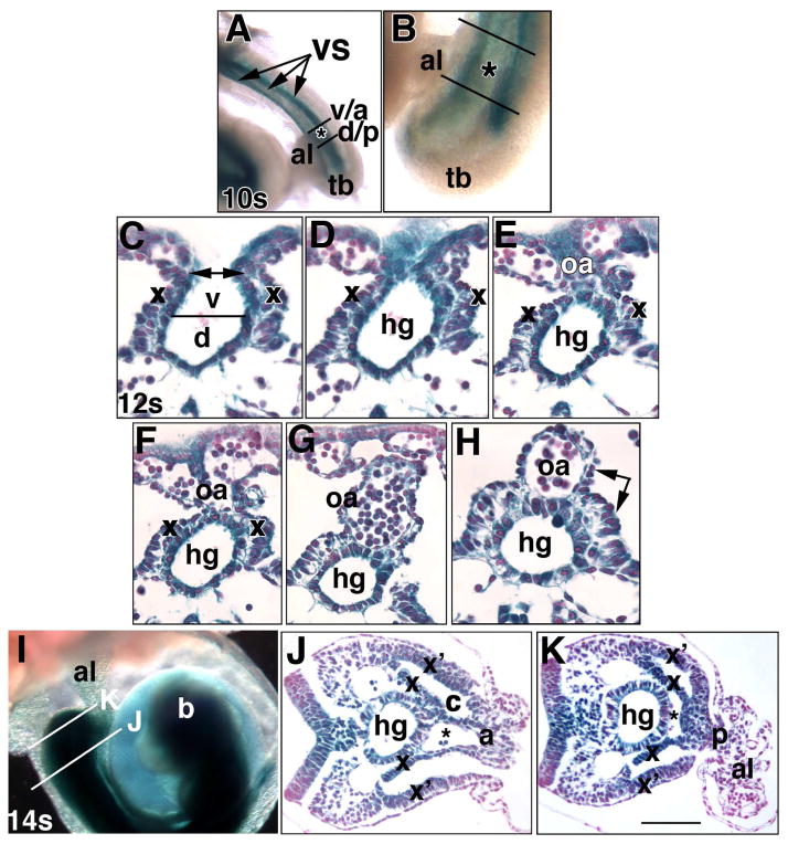
Fig. 5. Ptc-1 in the developing hindgut and omphalomeseneteric artery, proximal walls of the allantois, and the umbilical ring (Phase IIIa, ~8.75–9.25 dpc).
Specimens in all panels were prepared from the Ptc1:lacZ reporter mouse. Whole mount (A, B, I) and sectional (C-H, J, K) views. (A, B) 10-s stage (~8.75 dpc). Ventral views indicate the allantoic insertion into the ventral surface (VS, arrows, A, composed of the splanchno- and somatopleure). Black lines bracket the allantois (al) at the nascent umbilical ring; v/a: border of the insertion (asterisk) of the VCM/anterior umbilical wall into the VS; d/p: border of the insertion of the DCM/posterior umbilical wall into the VS. tb, tailbud. (C–H) Sequence of closure (C, double arrow) of the definitive endoderm (parsed into dorsal (d) and ventral (v) components, C) to form the hindgut (hg) tube (D–H) and omphalomeseneteric artery (oa) (E-H). x, splanchnopleure (C–I), double arrow, H, the splanchnopleure surrounds both the hindgut and OA. (I–K) 14-s stage (~9.25 dpc). The histological views displayed in J, K are indicated in (I) by white lines. b, developing brain. In J, K: x, splanchnopleure covering the hindgut; x’, lateral somatopleure. Together, x and x’ delineate the coelom (c). The vessel of confluence (asterisk) is continuous with the umbilical artery (J) at the level of VCM/anterior umbilical wall (a), and with the dorsal aortae (K) at the level of the DCM/posterior umbilical wall (p). Scale bar (K): 100 μm (J, K); 50 μm (C-H); 170 μm (B); 333 μm (I).

