Figure 3.
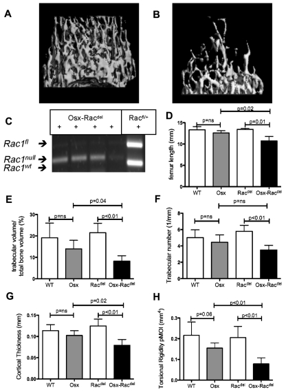
Osteoblast-restricted Rac deletion leads to defective bone acquisition in vivo. Micro-CT images of distal femoral trabecular bone architecture demonstrating normal bone structure in Racdel mice (Cre− controls; A) and marked reduction of trabecular bone volume and abnormal architecture in Osx-Racdel mice (B). (C) Validation of complete excision of Rac alleles in osteoblasts isolated by flow cytometry in OsxRacdel mice but not Osx-WT controls. (D) Bone length was reduced in Osx-Racdel mice (10.7 ± 1.1 mm) compared with Cre− controls (13.4 ± 0.3 mm, P = .01). No difference was observed between Osx and WT controls (12.6 ± 0.5 mm vs 13.3 ± 0.8 mm, respectively, P = .14; n = 3-5 biologic replicates for each genotype). (E) Trabecular bone volume was reduced in Osx-Racdel mice (8.2% ± 2.6%) compared with Cre− controls (21.5% ± 4.4%, P < .01). No difference was observed between Osx and WT controls (13.9% ± 4.1% vs 19.1% ± 6.9%, respectively, P = .15; n = 4-6 biologic replicates for each genotype; F) Trabecular number was reduced in Osx-Racdel mice (3.5 ± 0.6/mm) compared with Cre− controls (5.7 ± 0.8/mm, P < .01). No difference was observed between Osx and WT controls (4.4 ± 0.9/mm vs 5.0 ± 1.0/mm, respectively, P = .15; n = 4-6 biologic replicates for each genotype; G) Cortical bone thickness was reduced in Osx-Racdel mice (0.078 ± 0.014 mm) compared with Cre− controls (0.124 ± 0.016 mm, P < .01). No difference was observed between Osx and WT controls (0.102 ± 0.012 mm vs 0.113 ± 0.014 mm, respectively, P = .18). (n = 4-6 biologic replicates for each genotype; H.) Estimated cortical bone strength (as measured by the polar moment of inertia) was reduced in Osx-Racdel mice compared with Cre− controls (0.08 ± 0.03 mm4 vs 0.20 ± 0.05 mm4, P < .01). A trend to reduced cortical bone strength was observed in Osx transgenic controls compared with WT controls (0.15 ± 0.03 mm4 vs 0.22 ± 0.06 mm4, respectively, P = .06). (n = 4-6 biologic replicates for each genotype.) All values are means ± SD.

