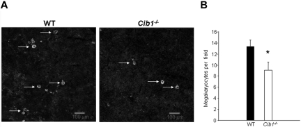
Figure 2.
Spleens of Cib1−/− mice contain fewer megakaryocytes than WT mice. (A) Fluorescent microscopic images of mouse spleen sections stained with anti-CD41 to label the megakaryocytes and Draq-5 to label nuclei. White arrows indicate megakaryocytes. Images were captured using a Zeiss 5-live confocal microscope (original magnification ×100). (B) Quantification of panel A expressed as megakaryocytes per field. *P < .05 vs WT (n = 7).

