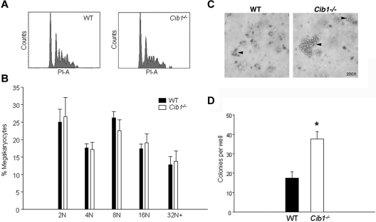
Figure 3.
CFU-MK production is heightened with Cib1 deletion. (A) Representative histograms of DNA content of CD41+ BM cells derived from WT and Cib1−/− mice. (B) Quantification of the ploidy distribution of WT and Cib1−/− BM megakaryocytes expressed as a percentage of total megakaryocytes (n = 6). (C) Representative light-microscopic images of CFU-MK stained with acetylcholinesterase after 7 days in culture. Arrows indicate CFU-MK colonies. (D) Quantification of the number of CFU-MK in each well after 12 days of incubation. Colonies were composed of at least 3 cells. *P < .05 vs WT (n = 4).

