
Figure 8.
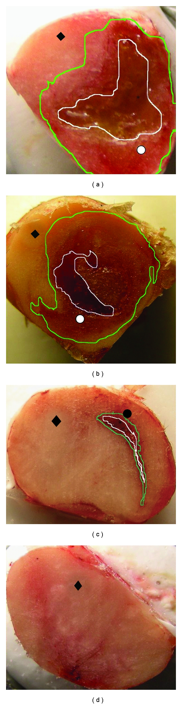
Macroscopic view of frozen tumor tissues at cryostat sectioning: (a) tumor treated with cisplatin/polymer formulation and excised 3 days after injection; (b) tumor treated with cisplatin/polymer formulation and excised 7 days after injection; (c) tumor treated with the blank polymer and excised 3 days after injection; (d) untreated tumor. The polymeric formulation is assigned with the star (∗), the necrotic tissue with the white circle (⚪), the infiltration of the inflammation cells with a black circle (•), and the intact tumor cells with a black rhomb (♦).