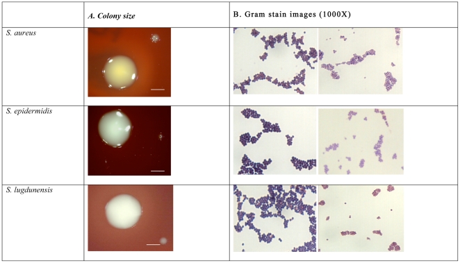
Figure 2. Morphological variations in size, pigmentation, haemolysis and Gram stain between WT and their corresponding SCVs in S. aureus, S. epidermidis and S. lugdunensis.
Column (A) shows differences in size and pigmentation between WT colonies (left) being larger and more pigmented than their SCVs (right) which are minute with diminished pigmentation (scale bar represents 1 mm). Column (B) shows the differences in response to Gram staining with WT cells (left) staining significantly darker than their corresponding SCVs (right).

