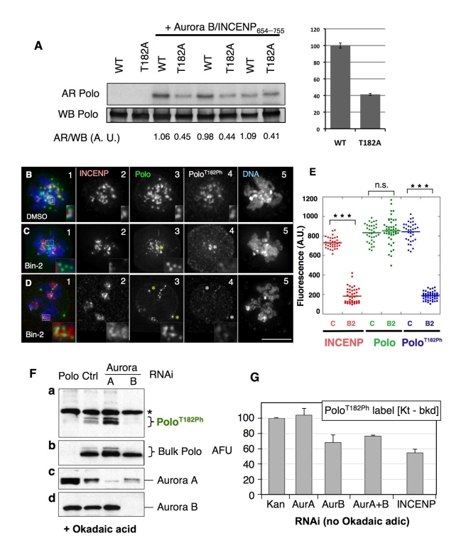
Figure 4. Aurora B activity is required for the activation of Polo kinase at the inner centromere.
(A) Aurora B phosphorylates Polo kinase in vitro. Bacterially expressed HIS-Polo or HIS-PoloT182A (which is catalytically inactive and therefore unable to autophosphorylate) were incubated with (or without) Drosophila Aurora B in complex with a fragment of INCENP (residues 654–755) in presence of 32P-g-ATP, in triplicate. Reaction products were resolved by SDS-PAGE transferred to nitrocellulose and analyzed by autoradiography (AR) and anti-Polo Western blot (WB). Quantitative measurements of signals were obtained (see Materials and Methods), and the ratios were calculated for each reaction (AR/WB, A.U.: arbitrary units). Right, average values for the relative phosphorylatin of PoloWT and PoloT182A by Aurora B. Error bars, SEM. (B–D) DMel-2 cells stably expressing Polo-GFP treated with (B) DMSO or (C–D) Binucleine-2, immunostained for INCENP, Polo, and PoloT182Ph (insets: zoomed images of kinetochores). In (C–D) asterisks indicate centrosomes. Merged images show INCENP/Polo/DNA. Zoomed images in (C–D) insets show examples of kinetochore pairs showing decreased levels of PoloT182Ph. (E) Dot plot showing the quantification of INCENP/Polo/PoloT182Ph signal intensity at the kinetochore (t test: *** p<0.0001; n.s., not significant; p = 0.4028). Signal intensities for individual kinetochores were measured using the SoftWorx Data Inspector tool; average background was subtracted; data was plotted using KaleidaGraph software. (F) RNAi depletion of Aurora B, but not Aurora A, strongly reduces PoloT182Ph levels in DMel-2 cells treated with okadaic acid. Cells were transfected with the indicated dsRNAs for 4 d, and 100 nM okadaic acid added for 4 h before immunoblotting to improve visualization of phosphorylated Polo. A dsRNA against the Kanamycin resistance bacterial gene was used as a negative control. Asterisks: non-specific bands. Both bulk Polo and PoloT182ph appear as doublets. (G) RNAi depletion of Aurora B or INCENP, but not Aurora A, reduces PoloT182Ph levels at centromeres/kinetochores. Cycling cells were treated with the indicated dsRNAs for 3 d (immunoblots are shown in Figure S6B) and PoloT182Ph was detected by immunofluorescence. Levels of PoloT182Ph at centromeres/kinetochores in prometaphase and metaphase cells were measured at individual kinetochores using Image J, subtracting background (Kt-bkd). Asterisks indicate centrosomes. Error bars = S.E.M.

