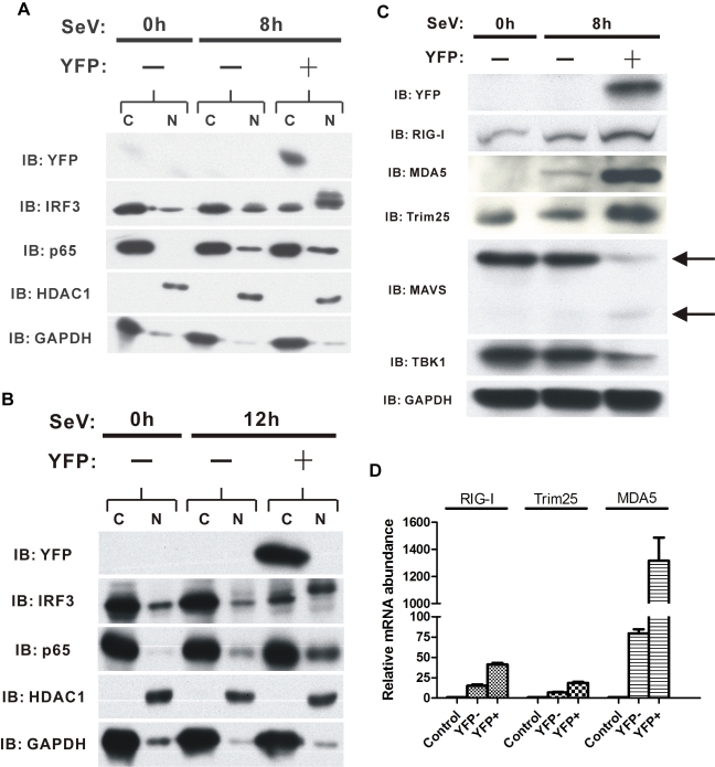
Figure 3. The RIG-I signaling pathway is activated in IFNβ-producing cells.
(A and B) Western blots showing cytoplasmic (C) versus nuclear (N) distribution of different factors present in FACS-sorted cells 8 h.p.i. (A) and 12 h.p.i (B). (C) Western blots showing cytoplasmic distribution of signaling pathway proteins present in FACS-sorted cells 8 h.p.i. Arrows indicate MAVS protein. (D) qPCR analysis illustrating the expression levels of RIG-I, Trim25, and MDA5 genes in sorted IFNβ/YFP homozygous MEF cells 8 h.p.i. IB, immunoblot.

