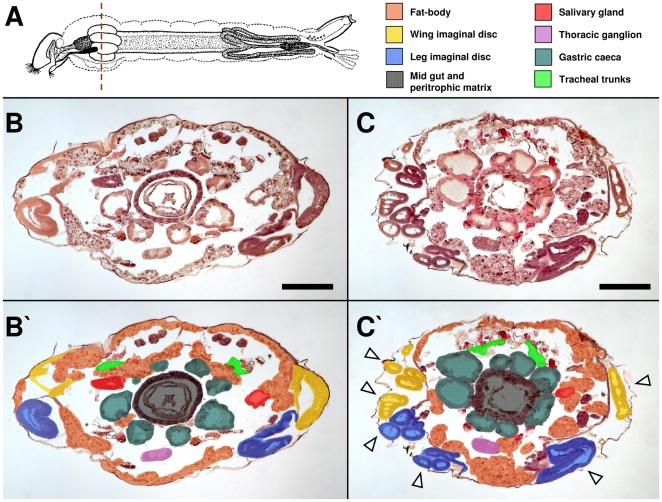
Figure 5. Novaluron induces histological alterations on Ae. aegypti larvae.
Bright field microscopy. (A) Larva scheme with histological section region evaluated (mesothorax, dashed line) and color caption for identified organs and tissues. HE staining of live L4l larvae from control (B, B′) and novaluron EI99 (C, C′) are shown. In (B′) and (C′) sections shown in the corresponding panels were colored to better identify structures. In (C′) arrowheads indicate disorganized imaginal discs (see Figure S2 for further details). In (A), larva scheme adapted from Christophers [3]. Bar = 200 µm.

