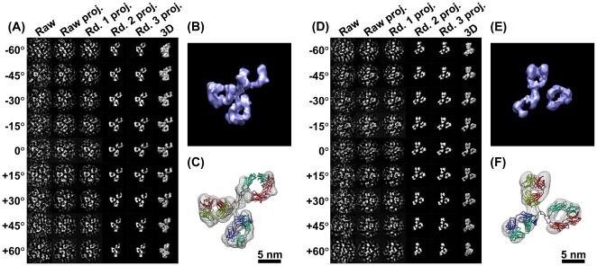
Figure 4. 3D reconstructions with negative staining of an IgG antibody by IPET.
(A) A single-instance of an IgG antibody was imaged by negative staining (NS) ET. Nine tilted views of the same single IgG particle were selected from 81 tilt micrographs that were CTF corrected by TOMOCTF, and then displayed in leftmost column. By FETR algorithm, the 3D reconstruction was iterated and converged. The corresponding tilting projections of the 3D reconstruction from major iterations were displayed beside the raw images in six columns. (B) The final 3D reconstruction of a targeted individual antibody particle was displayed in a contour level that contained a volume corresponding to 160 kDa. The reconstruction displayed three ring-shaped domains that corresponding to three domain of IgG antibody. (C) Docking the crystal structure (PDB entry 1IGT) of each domain of the IgG antibody into each ring-shaped density of IgG showed a good fit. The docking was performed by a rigid-body docking option in Chimera program [79]. The loops between the domains were simply connected by Chimera. (D) Another example of the IPET method in a 3D reconstruction of another single-instance of a targeted individual IgG antibody particle. The NS-ET images of the raw images and projections of the related reconstruction were also displayed. (E) The 3D reconstruction of this antibody particle displayed a similar structural feature to the first one, such as the three domains. (F) Docking the IgG antibody crystal structure into the density map showed a good fit as well.

