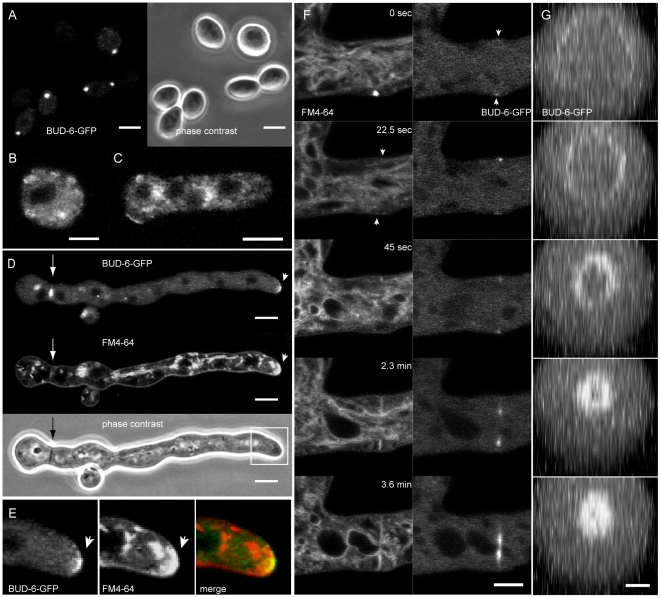
Figure 1. BUD-6 localization in conidia, conidial germlings and sites of septum formation.
(A) Intense and locally defined clusters of BUD-6 were localized at the cell poles of dormant macroconidia. Scale bars, 5 µm. (B–C) Specific recruitment of BUD-6 during isotropic expansion, cell symmetry breaking and germ tube outgrowth was not observed. Scale bars, 2.5 µm. (D) Localized recruitment of BUD-6-GFP to apical caps of growing germ tubes (arrowhead), as well as to septa (arrow), occurred in germlings ≥35 µm in length. FM4–64 stained vesicle clusters were observed at the same positions. (E) Enlarged view of the germ tube tip highlighted in (D). The merged image shows that BUD-6-GFP fluorescence only partially colocalizes with the FM4–64 stained apical cap which extends over a larger crecent. (F) Staining with FM4–64 revealed that BUD-6 recruitment to the incipient septation site preceded plasma membrane invagination (arrowheads), and that it constantly remained associated with the leading edge of the closing septum. Scale bar, 5 µm. See Movie S1 for time course sequences. Note: the bright FM-4–64 stained spot appearing at time point 0 sec does not colocalize with the cortical BUD-6 accumulation indicated by the arrowhead. (G) Reconstruction of BUD-6-GFP localization at the inner perimeter of the closing septal pore. Scale bar, 2.5 µm.

