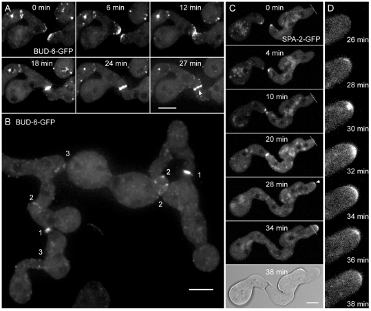
Figure 2. BUD-6 and SPA-2 recruitment during CAT-mediated cell fusion.
(A) Clusters of BUD-6-GFP became recruited to CAT tips, concentrated at the incipient fusion site upon contact, and formed an opening ring of fluorescence during fusion pore formation (arrowhead). See Movie S2 for time course sequences. Scale bar, 5 µm. (B) Typical changes in the BUD-6 localization pattern accompanied distinct stages of the cell-cell fusion process in germling networks: (1) pronounced accumulation shortly after CAT attachment, (2) ring formation during fusion pore opening, and (3) BUD-6 disappearance shortly after cytoplasmic continuity was established. Scale bar, 5 µm. (C) Z-projections of confocal optical sections through conidial germlings expressing SPA-2-GFP during CAT homing and fusion. Shortly after cytoplasmic continuity was successfully established and SPA-2 disappeared from the fusion site, a new cluster of SPA-2 became recruited to the germ tube tip (arrowhead) that resumed polarized growth (transient arrest of germ tube growth during cell fusion, and resumed tip extension post-fusion is indicated with dotted line). Scale bars, 5 µm. See Movie S3 for full time sequence. (D) Shows an enlarged view of BUD-6 recruitment during the continuation of germ tube tip growth as shown in (C).

