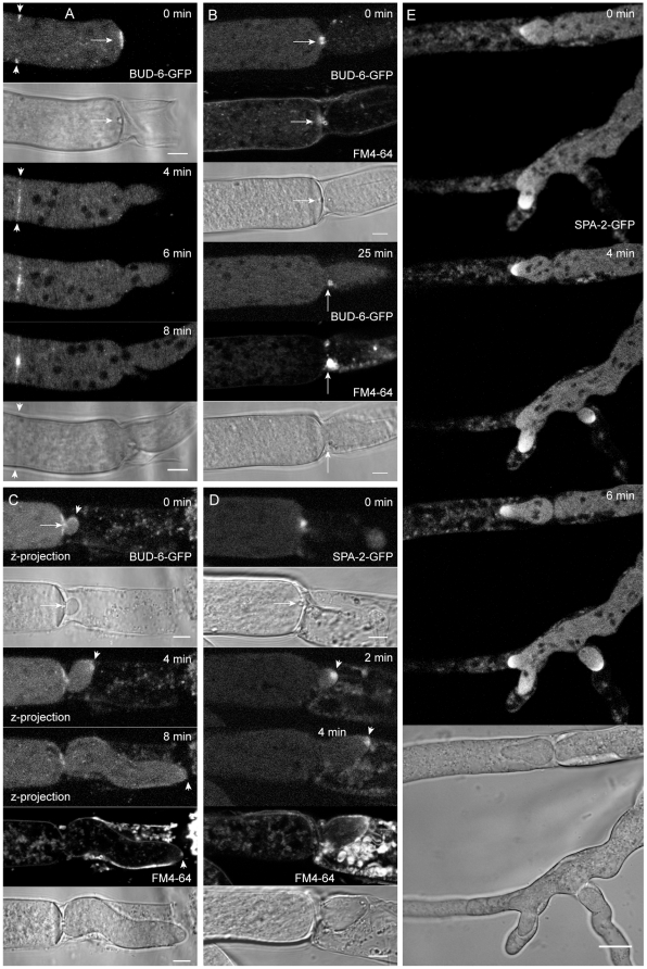
Figure 4. BUD-6 and SPA-2 concentrated at the septal plug prior to reinitiation of polarized tip growth.
(A) Upon physical injury a Woronin body occluded the septal pore which was surrounded by pre-existing BUD-6 fluorescence (arrow). At the same time, a new septum was being initiated 25 µm further back (arrowheads), again leaving BUD-6 at the septal pore after its completion. (B) At the onset of repolarization from the septum at the severed end of the hypha, BUD-6-GFP fluorescence became concentrated at the sealed pore, in the vicinity of the Woronin body and within an accumulation of lipophilic, possibly membranous, material stained by FM4–64 (arrows). See Movies S5 for full sequence. (C) The BUD-6 cluster often remained in place while the new tip emerged (arrowhead). Subsequently and coinciding with the condensation of a recognizable Spk, apical BUD-6 fluorescence became increasingly diffuse (arrowheads). See Movie S6 for full sequence. (D) SPA-2 rapidly became recruited to the occluded septal pore forming an intense spot associated with the Woronin body (arrow). The protein did not reside at consolidated septal pores, but rather relocated into an apical cap of the emerging tip (arrowhead) which accompanied extension of the new hyphae (E). See Movie S7 for full sequences of D. Scale bars A to D, 5 µm. Scale bar E, 10 µm.

