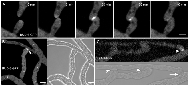
Figure 5. BUD-6 dynamics during vegetative hyphal fusion.
(A) BUD-6 became recruited to the tips of homing fusion hyphae, then concentrated at the attachment point and surrounded the opening fusion pore. Shortly after the pore was fully established BUD-6 fluorescence disappeared from this site. Scale bar, 5 µm. See Movie S8 for time course sequence. (B) Transient BUD-6 fluorescence accumulated at incipient fusion sites in the mature colony (arrowhead) and persistent BUD-6 signal at septal pores (all other fluorescently marked sites). Scale bar, 10 µm. (C) SPA-2-GFP became recruited to vegetative hyphal fusion sites (arrow). As it was never seen at completed fusion connections (arrowheads) it must follow the transient dynamics of BUD-6 in this context. Scale bar, 10 µm.

