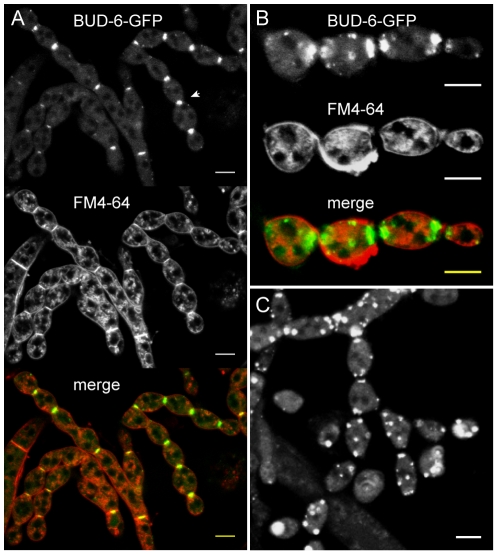
Figure 6. BUD-6 accumulation during conidiogenesis.
(A) BUD-6-GFP accumulated at septation sites in developing macroconidiophores (arrowhead). Scale bars, 10 µm. (B) In cytologically separated, but physically still attached conidia, BUD-6 fluorescence persisted at the cell poles; either at both or only at one pole in case of the terminal conidium. Scale bars, 10 µm. (C) In addition to strong fluorescence at the cell poles, bright clusters of BUD-6-GFP also accumulated at other locations of the cell cortex. Scale bar, 5 µm.

