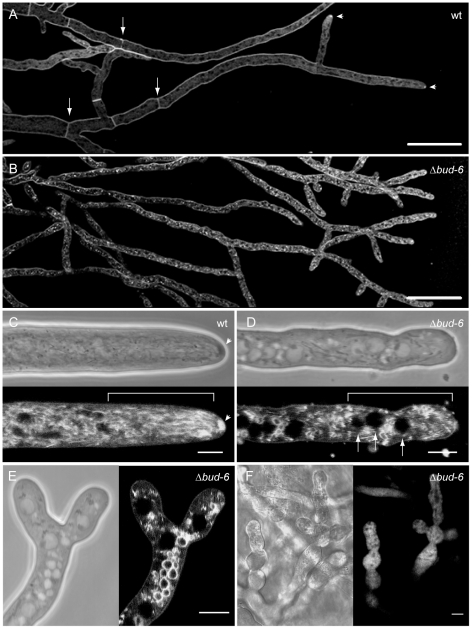
Figure 8. BUD-6 was required to organize the polarized growth apparatus at the hyphal tip.
(A and B) Comparison of FM4–64 staining pattern of mature hyphae at the leading edge of the colonies in the wild type and Δbud-6 mutant confirmed the absence of septa in the mutant (arrows in A indicate septa in the wt), as well as the absence of the Spk at the hyphal tips of Δbud-6 (arrowheads in A point toward wt Spk). Scale bars, 5 µm. (C and D) Close up of the apical and subapical area of polarized growing mature hyphae of wt and Δbud-6. The arrowheads in C indicate the Spk, which shows up as a dark sphere in the phase contrast image and was brightly stained by FM4–64. No such structure was observed in hyphae of the Δbud-6 mutant. The squared bracket marks the subapical nuclear exclusion zone in the wild type, which is not established in the Δbud-6 mutant. Here, nuclei (arrows) reach further up into the hyphal tip (also seen in E). Scale bars, 5 µm. (E) Apical branching and lack of hyphal tip organization in Δbud-6. Scale bar, 5 µm. (F) Immature and malformed conidiophores in Δbud-6. Scale bar, 10 µm.

