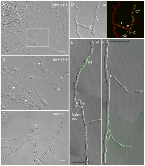
Figure 10. Δbni-1 and Δbud-6 strains displayed derepressed hyphal fusion at the colony edge.
(A) The lack of septa in Δbni-1 resulted in extensive accumulation of vacuoles at the leading edge of the colony. Scale bar, 50 µm. (B) Hyphal fusion at the leading edge of the colony, which is usually suppressed in the wild type through apical dominance, was observed in Δbni-1. Several fusion pores are indicated with arrowheads. Scale bar, 10 µm. See Movie S9 for time course sequence showing vacuolar passage through fusion connections. (C) After closer inspection, the same phenotype could be observed at the leading edge of Δbud-6 colonies. Scale bar 10 µm. (D–F) In the wild type, derepression of hyphal fusion at the leading edge occurred in the presence of conidial germlings. The establishment of cytoplasmic continuity – here visualized through the transfer of nuclei fluorescently labeled with histone H1-GFP (green) from conidial germlings into mature hyphae - involved either CAT-mediated cell fusion (D), or fusion pegs from the mature hypha (E and F) induced through the presence of conidial germlings. Arrowheads indicate fusion sites; C denotes the spore body; GT denotes the germ tube; H denotes mature hypha. Note that fluorescently labeled nuclei originating from the germling have only migrated into the upper part of the unlabeled wild type hypha. The arrowheads in (F) mark fusion pegs emerging from the mature hypha. Scale bar in D, 10 µm.

