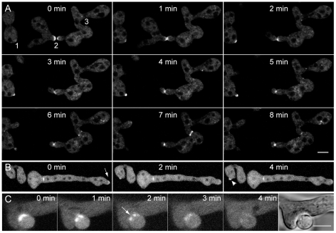
Figure 11. BNI-1 recruitment to sites of polarized growth, septum formation and cell fusion.
(A) In conidial germlings recruitment of fluorescently labeled BNI-1 was observed during three different cellular processes. (1) During cell symmetry breaking BNI-1 appeared at the cell cortex. (2) During CAT-mediated cell fusion, very bright accumulations of BNI-1 could be seen at the tips of interacting CATs. Due to a spore torque response upon cell-cell attachment, the left cell moved out of the focal plane. This movement is commonly observed when imaging germling fusion in liquid medium. (3) During septum formation BNI-1 was part of contractile actomyosin rings. Scale bar, 5 µm. (B) BNI-1 also accumulated in apical crescents at growing germ tube tips (arrow). Recruitment to septal pores and a new sites of cell symmetry breaking (arrowhead) showed up as well. Also note that BNI-1 is associated with septum formation (asterisk) at the base of the germ tube. (C) Consistent with its participation in CAT-mediated cell fusion, BNI-1 also became recruited to sites of vegetative hyphal fusion, and disappeared shortly after cytoplasmic continuity was established. The arrow marks the opening fusion pore. Scale bar, 5 µm.

