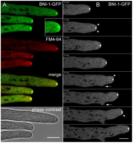
Figure 12. BNI-1 is a constituent of the Spk, but also localized to an apical cap.
(A) BNI-1-GFP colocalized with the Spk, but also was present in an apical cap (see inset in A). (B) Time sequence of BNI-1 dynamics during mature hyphal tip growth and lateral branch initiation. 0–2 min: in the straight growing hyphal tip small crescents (arrowheads) of BNI-1 were located on either side of the Spk. 4–6 min: shortly before branch initiation, tip extension transiently ceased – evident by rounding-off of the tip - and apical BNI-1 fluorescence disappeared. 8–10 min: extension of the main tip resumed with a brighter cluster of BNI-1 at the left hand side of the apex, followed by displacement of the Spk and left-orientation of the tip. A small crescent of formin fluorescence localized to the tip of the emerging branch (arrow). 12–14 min: a brighter cluster of the formin accumulated at the right hand side preceding reorientation of tip extension into this direction. Scale bars, 10 µm.

