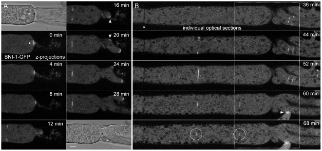
Figure 13. BNI-1 localization during septal plugging, tip repolarization and septum formation.
(A) Two minutes after damaging leading hyphae BNI-1 became recruited to the septal plug (position of the Woronin body is indicated with an arrow, 0 min). Fluorescence focused into a smaller area from which a new hyphal tip repolarized, and shortly after condensed into an subapical spot (arrowhead, 16 min) with flanking BNI-1 crescents on either side (inset, 20 min). In parallel, a new septum was being formed about 25 µm behind the severed end, and an additional site of polarity was established (arrowhead, 20 min). Scale bar, 5 µm. See Movie S11 for full sequence. (B) Continuation of (A) but with an extended field of view including an old septum (asterisk). The part of the hypha shown in (A) is outlined with a dashed box. A selection of individual optical slices shows the formation of several septa. Upon septum completion, BNI-1 gradually disappeared from the septal pore. Barely visible remains are indicated with circles at the 68 min time point. BNI-1 fluorescence was usually not observed at ‘old’ septa (asterisk). Recruitment of BNI-1 to a vegetative hyphal fusion site is indicated by an arrowhead.

