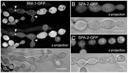
Figure 14. BNI-1 localization during conidiogenesis.
(A) During cytological compartmentalization of conidiophores BNI-1 localized to forming septa (arrowheads). Apart from occasional cortical clusters no specialized localizations (e.g. to cell poles) of the formin could be observed at mature stages of conidial development. (B) In contrast, apart from weak cytoplasmic fluorescence, no specific accumulation of SPA-2 could be observed during conidiophore formation, cytokinesis or (C) upon physical separation of mature conidia. Scale bars, 5 µm.

