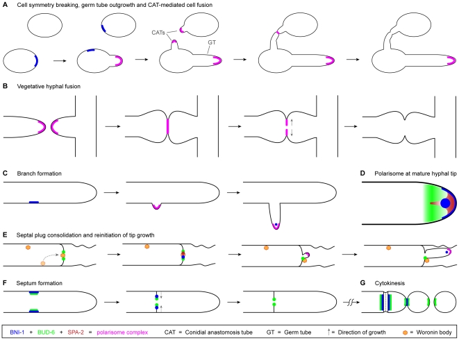
Figure 15. Schematic representation of the subcellular localization and dynamics of the three polarisome components SPA-2, BUD-6 and BNI-1 during key developmental stages of N. crassa.
(A) Prior to the constitution of the entire polarisome complex during germ tube growth and CAT-mediated cell fusion, BNI-1 accumulates at the incipient sites of cell symmetry breaking. (B) Equivalent to its dynamics during CAT fusion, the polarisome complex is present during homing and fusion of fusion hyphae, and removed once cytoplasmic continuity is achieved. (C) BNI-1 accumulates at the incipient site of branch formation, and together with BUD-6 and SPA-2, subsequently constitutes the complete polarisome complex as an apical crescent at the tip of the emerging branch. Finally, the polarisome adopts the mature hyphal tip configuration (D). (D) Configuration of the polarized tip growth apparatus in mature hyphae, including an apical cap and Spitzenkörper core of BNI-1, a fan-shaped distribution of SPA-2 inside the apical dome, and the subapical BUD-6 cloud. (E) Septal plugging and consolidation involves the Woronin body and all three polarisome proteins. During repolarization, a polarisome crescent is constituted at the emerging hyphal tip, and ultimately rearranged into its mature form (D). (F) Septum formation requires BNI-1 and BUD-6 – but not SPA-2 - as components of the CAR. Upon septum completion, only BUD-6 remains associated with the inner perimeter of the septal pore. (G) During cytokinesis BNI-1 and BUD-6 become recruited to the CAR and to forming secondary septa. Upon physical separation of macroconidia BNI-1 disappears, whereas BUD-6 remains at the cell poles. As during septum formation, SPA-2 has no role in this process.

