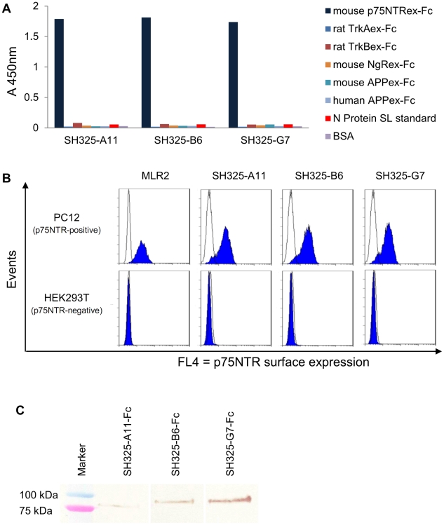
Figure 2. Characteration of the p75NTR-specific antibodies.
(A) Antigen binding ELISA. 100 ng of mouse p75NTRex-Fc (shown in dark blue), rat TrkAex-Fc, rat TrkBex-Fc, mouse NgRex-Fc, mouse or human APPex-Fc, N protein SL standard or BSA were immobilized in the plate for each well. 250 ng of each scFv was added after the antigen-coated plates were blocked with FCS for 1.5 hr. Bound scFvs were detected using anti myc-tag mAb (1∶500) and goat anti-mouse IgG HRP conjugated (1∶5,000). (B) The scFvs specifically recognize native p75NTR on PC12 cell surfaces. PC12 or HEK293T cells were stained with 250 ng of the p75NTR-specific scFvs (SH325-A11, SH325-B6, SH325-G7). Bound scFvs were detected by mouse anti-His6 mAb (1∶100) followed by goat anti-mouse IgG F(ab′)2 fragment APC conjugated (1∶200). The p75NTR surface expression on PC12 cells was determined by staining PC12 or HEK293T cells with mouse anti-p75NTR mAb (MLR2, 1∶200) followed by goat anti-mouse IgG F(ab′)2 fragment APC conjugated (1∶200). The blue histograms represent the mouse anti-p75NTR mAb (MLR2) or the p75NTR-specific scFvs staining PC12 cells (upper row) and HEK 293T cells (lower row). The white histograms represent the controls stained with goat anti-mouse IgG F(ab′)2 fragment APC conjugated alone (MLR2 line) or αphOx scFv (other lines) followed by mouse anti-His6 mAb and goat anti-mouse IgG F(ab′)2 fragment APC conjugated. (C) Detection of denatured antigen (p75NTRex-mFc) by the p75NTR-specific recombinant antibodies in immunoblot. 250 ng of p75NTRex-mFc was denatured and blotted on a PVDF membrane. 1 µg of each p75NTR-specific recombinant antibody was used to stain the membrane for 1.5 hr. The bound antibodies were detected by goat anti-human IgG Fc antiserum AP conjugated (1∶2,000) for 1 hr at RT.

