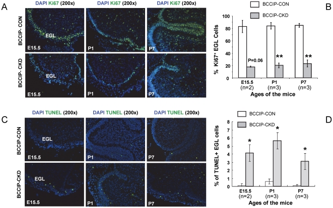
Figure 7. proliferation defects and excessive cell death in the external germinal layer (EGL) granule cell progenitors.
Ki67 IHC staining was used to identify the proliferative cells (Green in panel A) and TUNEL assay was performed to identify the apoptotic cells (Green in panel C). DAPI staining (Blue) was used to identify the nuclei of the cells. (A) Ki67 staining positive proliferative cells. (B) Quantification of Ki67 staining. (C) Apoptotic cells in the EGL at age E15.5, P1, and P7. (D) Quantification of TUNEL staining. The “n” values indicate the pairs of littermate matched mice used in the assay. Data are averages and standard errors from the indicated number of mice. *: P<0.05; **: P<0.01; ***: P<0.001.

