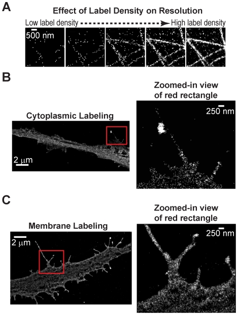
Figure 1. Comparison between cytoplasmic and membrane labeling for neuron imaging.
(A) STORM images of microtubules demonstrating the effect of label density. In the first panel the localizations from only the first few hundred frames of a STORM movie are included in the reconstructed image to simulate the effect that would be observed in the case of low label density. In the last panel localizations coming from the entire STORM acquisition are included to simulate the effect that would be observed in the case of high label density. The panels in between include progressively increasing number of localizations in the final reconstructed image. It is not possible to reconstruct the actual microtubule structure from the first image due to the low number of localizations, whereas the ability to reconstruct the microtubule structure increases with increasing number of localizations. (B) 2D STORM image of a neural process expressing YFP in the cytoplasm. The YFP was immuno-labeled with antibodies conjugated to photoswitchable A405-A647 pair for STORM imaging. The zoomed-in view shows a region with small neural processes. The small volume of these processes results in a low localization density in STORM images. (C) 2D STORM image of a neural process expressing mCherry attached to the membrane through a palmitoylation sequence. The mCherry was similarly immuno-labeled with antibodies conjugated to photoswitchable A405-A647 pair. The zoomed-in view shows a region of small neural processes. The membrane targeting resulted in a 3.6-fold improvement in label density.

