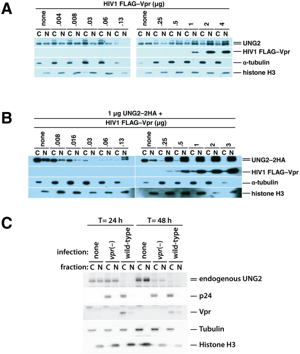
Figure 4. HIV1 Vpr-mediated degradation and subcellular redistribution of UNG2 are dose-dependent.
293T HEK cell cultures were transfected with empty vector alone and together with increasing quantities of HIV1 FLAG–Vpr expression vector as indicated. 48 hours after transfection nuclear and cytoplasmic fractions were prepared and analyzed by immunoblotting with antibodies specific for UNG2, FLAG epitope tag (FLAG–Vpr), α tubulin (cytoplasmic fraction control) and Histone H3 (nuclear fraction control) (A). 293T cultures were transfected with 1 µg of UNG2–HA expression vector alone, together with increasing quantities of HIV1 FLAG–Vpr expression vector as indicated. 48 hours after transfection nuclear and cytoplasmic fractions were prepared. Immunoblotting with anti-HA (UNG2–2HA), anti-FLAG (FLAG–Vpr), anti-α tubulin (cytoplasmic fraction control) and anti-Histone H3 (nuclear fraction control) antibodies was used to determine relative quantities of the respective protein that were present in the fractions (B). HEK 293T cells, either untransfected (left) or transfected with 1 µg of UNG2–2HA expression vector and 3 µg of empty vector (C, right) were mock-infected (none), infected with vpr(–), env(–), VSV-G-pseudotyped virus (vpr(–)) or with wild-type, env(–), VSV-G-pseudotyped virus (wild-type).

