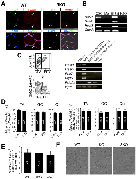
Fig. 1.
Expression of Hesr family genes and the Hesr1-null and Hesr3-null skeletal muscle phenotypes. (A) Transverse sections of tibialis anterior muscle from wild-type (WT) and Hesr3-null (3KO) mice (a negative control for anti-Hesr3 antibody) were stained with antibodies to laminin α2 (violet), Pax7 (green) and Hesr3 (red) and with DAPI (blue). Arrowheads indicate Pax7-expressing cells lying beneath the basal lamina. (B) RT-PCR of Hesr family genes in quiescent satellite cells (QSC) and myoblasts [Mb; cultured for 3 days in growth medium (GM)]. A whole E13.5 embryo and water were used as positive and negative controls, respectively. (C) RT-PCR of Hesr1 and Hesr3 in mononuclear cells derived from 10-week-old uninjured skeletal muscle. FACS profiles show each cell population used in RT-PCR. (D) Tibialis anterior (TA), gastrocnemius (GC) and quadriceps (Qu) muscle weight (mg) per gram body weight of 3-month-old male Hesr1-null (1KO), 9-month-old male 3KO and control littermate (cont) mice. The y-axis shows the mean with s.d. (E) The number of Pax7+ satellite cells in 10-week-old female uninjured TA muscle of WT, 1KO and 3KO mice. The y-axis shows the mean number of satellite cells per 100 cross-sectional myofibers with s.d. (F) A TA muscle of each 8-week-old mouse was injected with cardiotoxin and the muscles were fixed 2 weeks after the injection. The number of mice used in each study is indicated. Scale bars: 25 μm in A; 100 μm in F.

