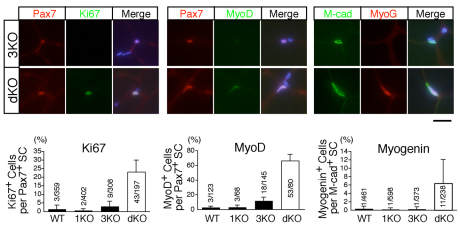
Fig. 3.
Myogenic and proliferative marker expression in adult dKO satellite cells. Uninjured TA muscles of 10-week-old female dKO and 3KO mice were stained with Ki67 (green), MyoD (green) and myogenin (red) antibodies. Pax7 (red) and M-cadherin (green) antibodies were used to detect satellite cells. Scale bar: 20 μm. The graphs beneath indicate the frequency of each marker-positive cell in WT, 1KO, 3KO and dKO mice. The y-axis shows the mean value with s.d. (n=3-5). The number of marker-positive satellite cells among total counted satellite cells is indicated in each bar.

