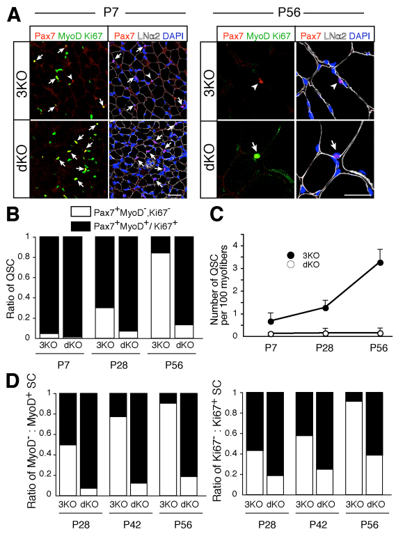
Fig. 4.
Failure of dKO satellite cells to enter the undifferentiated quiescent state. (A) Quantitative analysis of undifferentiated quiescent satellite cells in uninjured TA muscle at P7 and P56. Arrows and arrowheads indicate differentiated non-quiescent (Pax7+, Ki67+/MyoD+) and undifferentiated quiescent (Pax7+, Ki67–, MyoD–) satellite cells, respectively. Pax7, red; MyoD/Ki67, green; laminin α2, white; DAPI, blue. Scale bars: 25 μm. (B) Ratio of undifferentiated quiescent (white) to differentiated non-quiescent (black) satellite cells in uninjured TA muscle of dKO and littermate 3KO mice at the indicated ages. The value shown is an average of the results of experiments conducted with three to four mice. (C) The number of undifferentiated quiescent satellite cells in B. The y-axis shows the mean number of satellite cells per 100 cross-sectional myofibers with s.d. (D) Relative ratio of MyoD– (white) to MyoD+ satellite cells (black) and Ki67– (white) versus Ki67+ satellite cells (black). The value shown is an average of the results of experiments conducted with three mice. The x-axis indicates the age of the mice analyzed.

