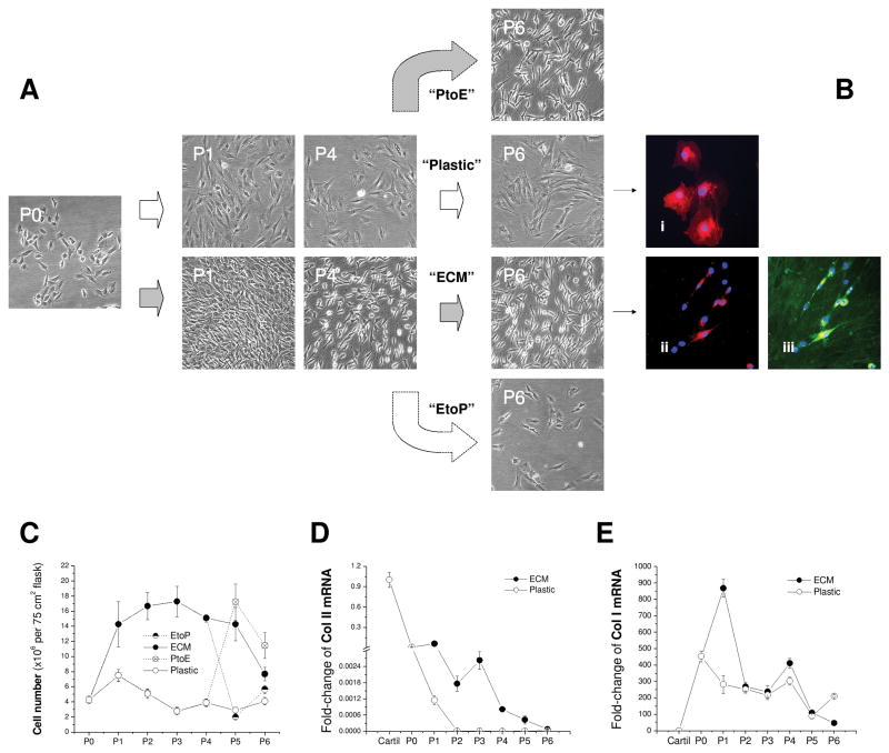
Fig. 1.
In vitro expansion of articular chondrocytes on Plastic, ECM, PtoE, or EtoP from P0 to P6. (A) Representative cell morphology from chondrocytes at P0, P1, P4, and P6. (B) Dil-stained chondrocytes (red) with DAPI as a nuclear counterstain (blue) were expanded on either Plastic (Bi) or ECM [immunostained without (Bii) or with (Biii) collagen I antibody (green)]. (C) Expanded cell number was compared for plating on either ECM or Plastic at each passage. (D) Fold-change of collagen II (Col II) mRNA by the mRNA value from native cartilage was compared in cartilage tissue and chondrocytes expanded on ECM or Plastic. (E) Fold-change of collagen I (Col I) mRNA by the mRNA value from native cartilage was compared in cartilage tissue and chondrocytes expanded on ECM or Plastic. Data are shown as average ± SD for n = 4.

