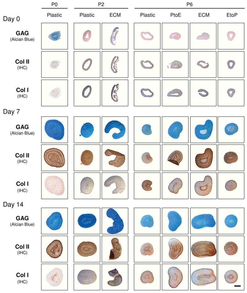
Fig. 3.
Redifferentiation capacity of expanded chondrocytes was assessed at the protein level using histology after chondrocyte-pellets were incubated in a TGF-β1-containing medium for 14 days. Alcian blue staining was for sulfated GAGs with fast red as a counterstain, and immunohistochemistry (IHC) was for collagens I and II (DAB substrate had a brown color suggestive of positive staining) with hematoxylin as a counterstain. The scale bar was 800 μm.

