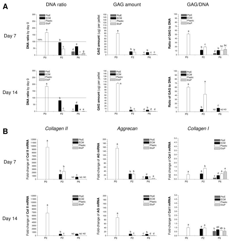
Fig. 8.
Redifferentiation capacity of expanded chondrocytes was assessed at the protein and mRNA level using chondrocyte-pellets after incubation in a serum-containing medium for 14 days. (A) Biochemical analysis was used to quantitatively measure DNA and GAG amounts in the 7- and 14-day pellets. DNA data were represented by DNA ratio adjusted by DNA amount at day 0. Ratio of GAG to DNA was used as a chondrogenic index. (B) TaqMan® RT-PCR was used to quantitatively measure chondrogenic marker genes (collagen II, aggrecan, and collagen I) in the pellets of expanded chondrocytes at days 7 and 14. Data are shown as average ± SD for n = 5. Groups not connected by the same letter are significantly different (p<0.05).

