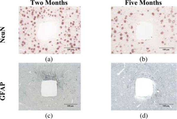
Fig. 10.
Histologic sections of rabbit cerebral cortical tissues showing tissue response at different times after implantation. (a) and (b) NeuN stain for neurons. (c) and (d) GFAP stain for astrocytes and their processes. The tissues were sectioned perpendicular to the length of probe shanks after the arrays were removed from the tissue. (a) and (c) Two months after implantation. Astrogliosis is visible mostly near one side of the track. (b) and (d) Five months after implantation. Astrogliosis is reduced from that occurring at two months.

