
Fig. 6. Changes in tuning curve, best frequency and response magnitude of thalamic and collicular DSCF neurons evoked by focal activation or inactivation of cortical DSCF neurons in the mustached bat, Pteronotus parnellii parnellii.
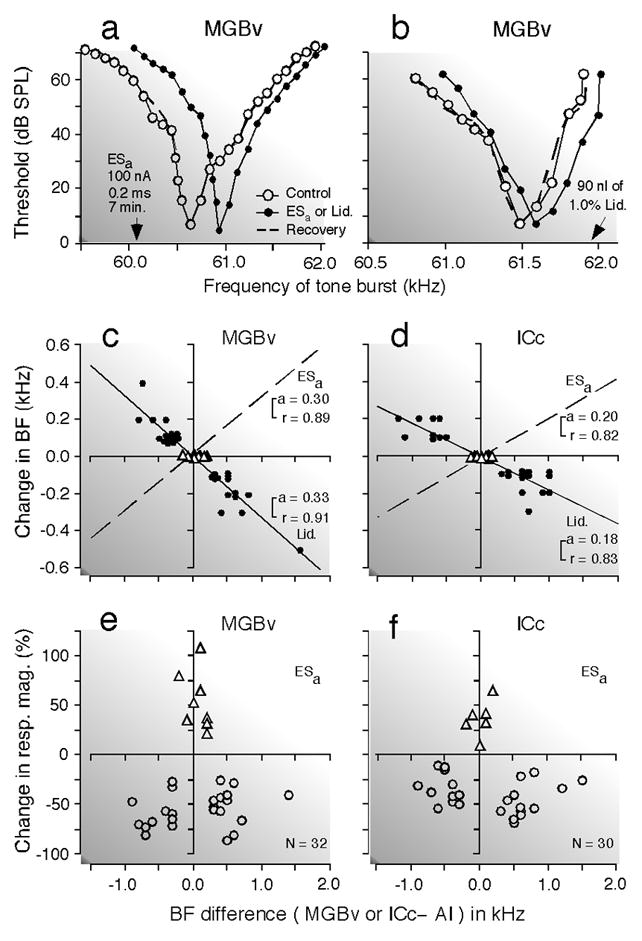
(a) and (b) Shifts in the frequency-tuning curves of two thalamic (MGBv) neurons evoked by an activation (a) or inactivation (b) of cortical neurons: activation by electric stimulation of a 0.2 ms, 100 nA electric pulse delivered at a rate of 5/s for 7 min (ESa) and inactivation by 90 nl of 1.0% lidocaine (Lid.). The best frequencies (BFs) of the activated or inactivated cortical neurons are indicated by the arrows. The curves were measured before (control, open circles), during (closed circles), and after (recovery; dashed lines) the cortical activation or inactivation. The data points for the recovery are not shown because almost all of them overlapped with those for the control. (c) and (d) The BF shifts of thalamic (c) and collicular (d) neurons evoked by a focal activation (dashed lines) or inactivation (solid lines and filled circles) of cortical neurons. The abscissae represent the differences in BF between the stimulated cortical (AI) and recorded thalamic (MGBv) or collicular (ICc) neurons in the control condition. The abscissae are the same as those in (e) and (f). The BFs of the stimulated cortical neurons were 61.2 kHz on the average. The triangles and circles represent the data obtained from matched and unmatched subcortical neurons, respectively. The regression lines, their slopes ‘a’ and correlation coefficients ‘r’ are shown in the graphs. The BF shift is centrifugal for the cortical activation, but centripetal for the cortical inactivation. (e) and (f). The ordinates represent the percent change in the response magnitude (number of pulses per tone burst) of thalamic (e) and collicular (f) neurons evoked by the cortical activation. The triangles and circles, respectively, represent percent changes in the response magnitude of matched and unmatched subcortical neurons at the BFs of individual neurons in the control condition. To measure response magnitudes, tone bursts were set at the best amplitude of each neuron in the control condition. Changes in BF (c and d) and response magnitude (e and f) both are larger in the MGBv than in the ICc (Zhang and Suga 2000).