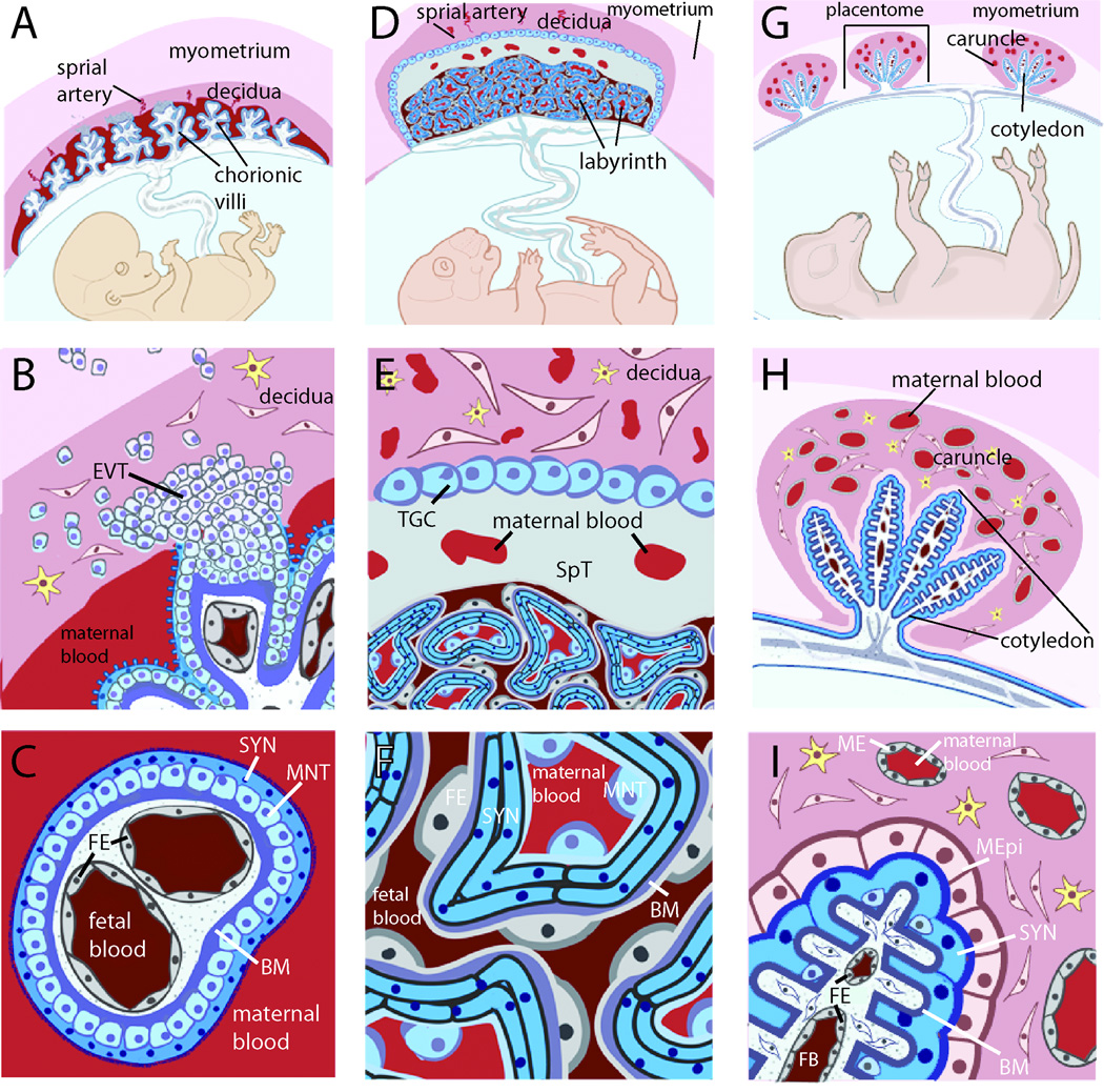
Fig. 1. Placental structures are diverse and do not necessarily cluster evolutionarily, complicating the selection of appropriate model systems.
A. The hemomonochorial human placenta is a villous structure bathed in maternal blood and anchored deep in the uterine decidua by extravillous trophoblasts. B. The human uterine-trophoblast interface is composed of decidual fibroblasts and maternal leukocytes juxtaposed with fetal extravillous trophoblasts (EVT), some of which migrate into the myometrium, while others remodel maternal spiral arteries (not shown). Many remain connected to a column of trophoblasts that extends into the fetal villus and stops at a basement membrane that divides trophoblasts from fetal capillaries. C. The human blood-trophoblast interface, showing villus cross-section. Maternal blood is separated from fetal vessels by a single syncytiotrophoblast (SYN), whose apical surface is covered by dense branched microvilli. Underlying that is a layer of mononuclear subsyncytial trophoblasts (MNT) that is continuous in early pregnancy but semi-discontinuous in later trimesters. Both structures are undergirded by a basement membrane. D. The hemotrichorial mouse placenta places maternal and fetal vessels in close contact within a nutrient-exchange labyrinth that is anchored in the decidua E. via a spongiotrophoblast (SpT) region and trophoblast giant cells (TGC) (analogous to EVT). The decidua is primarily composed of leukocytes and fibroblasts, as well as maternal arteries modified by trophoblast giant cells (not shown). F. Cross-section of labyrinthine region. From the maternal blood, a semi-discontinuous layer of mononuclear trophoblasts (MNT) overlays two layers of synctiotrophoblast with limited, non-overlapping cell-cell junctions. Beyond this lies fetal endothelium (FE). G. The sheep placenta is primarily epitheliochorial, and composed of multiple placentomes throughout the uterus. H. Each placentome includes a uterine caruncle rich in endometrial glands (not shown) and partially encapsulating a relatively non-invasive fetal cotyledon. I. Much of the cotyledonary surface effaces with a maternal epithelium (MEpi; which is not fully continuous and considered synepitheliochorial in regions, not shown). Maternal blood is separated from fetal blood by maternal endothelium (ME), endometrial fibroblasts, maternal epithelium, fetal syncytiotrophoblast (SYN), chorionic fibroblasts and fetal endothelium (FE). B–I. yellow cells = maternal leukocytes. A-F. Adapted from [5].

