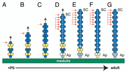
Figure 8.
Hypothetical formation of a blue (β-gal-positive) stripe spanning the whole adult adrenal cortex by a combination of edge-biased growth and centripetal cell displacement. Diagram showing initial edge-biased growth (A–C) followed by transition (D) to stem cell maintenance (E–G) in the adrenal cortex. (A–C) show the production of a blue (β-gal-positive) stripe in the outer adrenal cortex, driven by the onset of edge-biased growth (as in Fig. 7A), leaving a mosaic region in the inner cortex (perhaps in the X zone). The horizontal arrows indicate the proliferating cell(s), and the vertical black arrows indicate increased radial growth. (E–G) show extension of the blue stripe toward the medulla by production of new cells in the outer cortex, balanced by loss of cells in the inner cortex following the onset of tissue maintenance. Activated stem cells in the outer cortex (single horizontal arrow) self-renew, produce more differentiated daughter cells (probably equivalent to transient amplifying cells), which move centripetally, divide in the outer third of the cortex and displace existing cells toward the medulla, where they finally die by apoptosis (indicated by stippled shading of the bottom three cells). The grouped horizontal arrows represent the region of the cortex where cells proliferate (see text). The white arrow shows the direction of cell displacement and points to the same cell as it is displaced centripetally until it undergoes apoptosis. This process erodes the original mosaic pattern remaining in the inner cortex and replaces it with a continuous blue stripe, which spans the whole cortex. (D) shows a hypothetical transition stage between adrenal growth and maintenance, where the adrenal cortex is still growing, but stem cells have become activated in the outer cortex. some daughter cells also divide in the outer half of the cortex (so broadening the proliferative zone to include the ZG and the outer ZF), and cell death begins in the inner cortex, adjacent to the medulla. Ap, apoptosis (cell death); SC, stem cell. In greyscale prints of the figure, blue hexagons appear dark, and yellow hexagons appear light.

