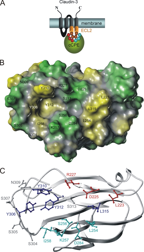
FIGURE 1.
Putative binding pocket for claudins on surface of cCPE structure (PDB 2QUO (18)). A, scheme of cCPE (green) binding to ECL2 (orange) of Cld3 or Cld4. Residues investigated are shown as circles (red and cyan, in cCPE; orange, in ECL2). B, hydrophobic potential calculated for surface of cCPE structure showing polar (green) and hydrophobic (yellow) areas for residues (labeled) that surround putative binding pocket for claudins. C, cCPE structure showing positions of residues (ball and stick) in more detail (blue, previously described positions for which Ala substitution inhibits binding; gray, previously described positions for which Ala substitution increases binding (16); red/cyan, reported in this study).

