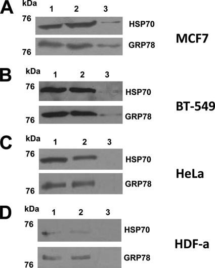
FIGURE 5.
WB of the affinity-purified TDF-R candidates. The TDF-R candidates were purified by AP using TDF-P1 peptide, and the eluate was investigated by WB using anti-GRP78 and anti-HSP70 antibodies. The cell lysate was prepared from MCF7 steroid-responsive cells (A), BT-549 steroid-resistant cells (B), HeLa cancer cells (C), and HDF-a cells (D). The molecular mass marker is shown (in kDa) on the left. Each WB contains input cell lysate (lane 1), flow-through (lane 2), and eluate (lane 3) of the AP experiments.

