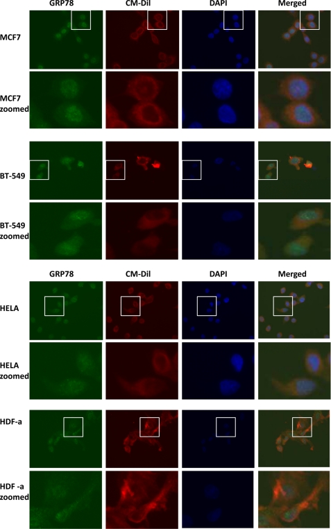
FIGURE 6.
IF detection of GRP78 protein. Expression of GRP78 was investigated in steroid-responsive MCF7 and steroid-resistant BT-549 breast cancer cells, non-breast HeLa cancer cells and normal HDF-a. The cells were incubated with anti-GRP78 antibodies and then AlexaFluor 488 antibodies for detection of GRP78 protein (green). Plasma membrane was stained with CM-Dil (red), and nuclei were stained with DAPI (blue). The merged images are also shown. Enlarged images for each cell line are also shown.

