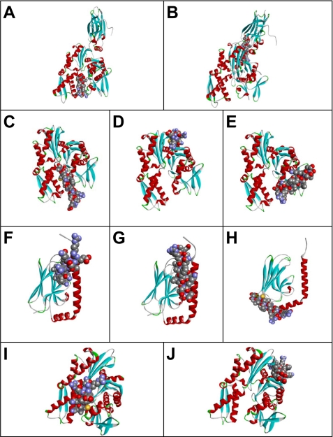
FIGURE 9.
Possible peptide binding pockets tentatively identified by GRAMM-X Protein-Protein Docking Web Server version 1.2.0. The TDF-P1 peptide (P1) binding pockets were predicted using Protein Data Bank structures 1YUW (A and B), 3LDN (C–E), 3N8E (F–H), and 2E88 (I and J). Receptor proteins are shown in the form of a ribbon diagram colored by secondary structure. P1 peptide is shown in space-filling mode colored by atom type.

