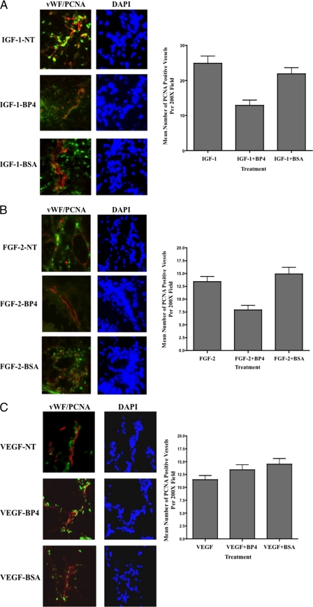
FIGURE 3.
Reduced blood vessel proliferation associated with IGFBP-4-mediated inhibition of angiogenesis. CAM tissues from each experimental condition were harvested and snap frozen. 4-μm tissue sections were analyzed by immunofluorescence co-staining. CAM tissues stimulated with IGF-1 (A), FGF-2 (B), and VEGF (C) were co-stained for expression of vWF to mark blood vessels (red) and PCNA (green) to mark proliferating cells. Left panels, representative examples of CAMs co-stained for vWF and PCNA from each experimental condition. Right panels, quantification of the number of PCNA-positive proliferating blood vessels per 200× microscopic field for each experimental condition. Data bars represent the mean number of proliferating blood vessels (± S.E.) from five 200× fields from each of four different CAMs per condition.

