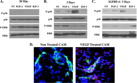
FIGURE 4.
Elevated levels of phosphorylated p38 MAPK detected in VEGF-stimulated angiogenic CAM tissue. To examine changes in expression of MAPKs within angiogenic CAM tissues, lysates were prepared from pooled CAM tissues (n = 4) from each experimental condition. Equal amounts of CAM lysates from each experimental condition were analyzed for the relative levels of p38 and ERK. A, Western blot of untreated CAMs (NT) and CAMs stimulated for 30 min with FGF-2, VEGF, and IGF-1 and analyzed for the expression of total and phosphorylated p38 and ERK. B, Western blot of untreated CAMs (NT) and CAMs stimulated for 3 days with FGF-2, VEGF, and IGF-1 and analyzed for the expression of total and phosphorylated p38 and ERK. C, Western blot of untreated CAMs (NT) and CAMs stimulated for 3 days with FGF-2, VEGF, and IGF-1 in the presence of IGFBP-4 and analyzed for the expression of total and phosphorylated p38 and ERK. D, representative examples of CAMs (n = 4) co-stained for vWF (green) and phosphorylated p38 (red) from untreated and VEGF-treated CAMs. Photos were taken at 400× magnification.

