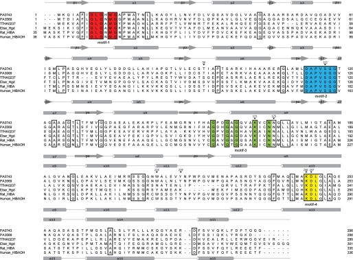
FIGURE 1.
Structure-based sequence alignment of PA0743 and several β-hydroxyacid dehydrogenases. The secondary structure elements of PA0743 and human HIBA dehydrogenase are shown above and below the alignment, respectively. Residues conserved in all aligned β-hydroxyacid dehydrogenases are boxed. The residues comprising the four characteristic β-hydroxyacid dehydrogenase sequence motifs (3) are highlighted in different colors and labeled below the alignment. The PA0743 residues mutated to Ala in this work are marked with an asterisk above the alignment and numbered. The proteins compared are PA0743 (UniProTKB Q9I5I6), PA3569 (P28811), rat HIBA dehydrogenase (P29266), the HIBA dehydrogenase TTHA0237 from T. thermophilus (Q5SLQ6), 2-(hydroxymethyl)glutarate dehydrogenase Hgd from E. barkeri (Ebar) (Q0QLF5), and the predicted human HIBA dehydrogenase (HIBADH) (P31937).

