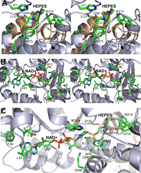
FIGURE 6.
Close-up stereoview of PA0743 active site. A, substrate-binding site with the modeled HEPES molecule. B, cofactor-binding site with the bound NAD+. C, the relative orientation of NAD+ and HEPES near the catalytic Lys-171 in the active site. Two PA0743 structures (Protein Data Bank codes 3OBB and 3Q3C) were superimposed, and the PA0743 ribbon from 3OBB is shown with the bound NAD+ and HEPES. The amino acid side chains and ligands are shown as sticks and labeled along with the protein ribbon of two protomers colored in gray and tan.

