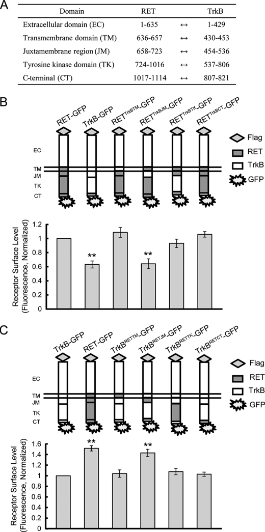
FIGURE 4.
RET juxtamembrane domain is responsible for the differential RET and TrkB surface levels. A, amino acid position of each domain based on published sequences is shown. B, surface levels of chimeric RET-GFP receptors with domain substitutions of TrkB were measured using a ratiometric fluorescence assay in transfected PC12 cells. Schematic diagrams show the structures of RET (gray) and TrkB (white) subdomains. Relative surface expression levels of each chimera were normalized to that of RET-GFP. The results are represented as mean ± S.E. from three independent experiments (**, p < 0.01 versus RET surface levels; one-way ANOVA). C, surface levels of chimeric TrkB receptors with domain substitution of RET were determined as in B. Schematic diagrams show the structures of TrkB (white) and RET (gray) subdomains. Relative surface expression levels of each chimera were normalized to that of TrkB-GFP. The results are represented as mean ± S.E. from three independent experiments (**, p < 0.01 versus TrkB surface levels; one-way ANOVA).

