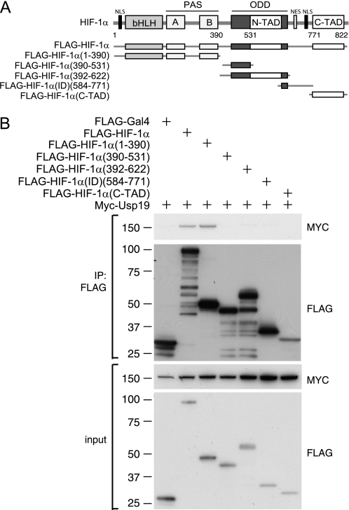
FIGURE 2.
Mapping HIF-1α interaction domain. A, schematic illustration of truncated, FLAG-tagged, HIF-1α constructs. bHLH, PAS domain, ODD, N/C-terminal transactivation domain (N-TAD and C-TAD), nuclear localization signal (NLS), and nuclear export signal (NES) are indicated. B, co-immunoprecipitations (IP) using a FLAG(M2) affinity gel from lysates of HEK293T cells co-transfected with the truncated forms of HIF-1α or FLAG-GAL4 as control, together with Myc-USP19.

