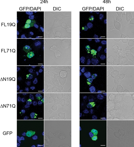
FIGURE 1.
Cellular localization of the ATN1 variants. GFP-tagged FL-ATN1 variants partitioned between the cytoplasm and the nucleus and ΔN-ATN1 variants were localized in the cytoplasm. The nuclear localization was confirmed with DAPI counterstaining. N2a/Tet-Off cells were transiently transfected with GFP-tagged ATN1 variants and visualized by fluorescence (GFP) and differential interference-contrast (DIC) microscopy after 24 and 48 h. GFP was soluble over the whole expression cycle and is included as a control. Scale bar = 10 μm.

