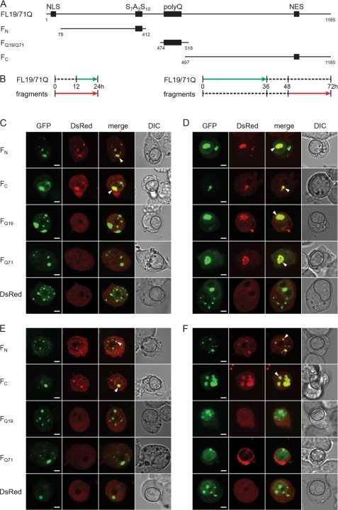
FIGURE 4.
Orthogonal cross-seeding in vivo reveals polymorphic interactions of nuclear FL19Q and FL71Q inclusions with the ATN1 fragments. A, schematic of the ATN1 fragments used in the cross-seeding experiments. The numbering is according to the amino acid sequence of FL19Q. B, expression scheme for the coexpression of the fragments and FL-ATN1. Each expression scheme is representative for the image series below it. C and D, recruitment properties of the FL71Q aggregates at early (C) and late (D) expression times. E and F, recruitment ability of FL19Q inclusions at early (E) and late (F) times of expression. N2a/Tet-Off/LacI cells were cotransfected with GFP-tagged FL19Q or FL71Q and a DsRed-tagged fragment thereof (marked on the left of each image row). The degree of colocalization is illustrated by merging the GFP and DsRed images. The recruited fragments in the FL-ATN1 inclusions are marked with an arrow on the merged images. The boundaries of the nuclei are designated with a black line on the differential interference contrast (DIC) images. Note that there are some hyperfluorescent spots on the FQ19 (D) and FC (F) images (visible on the DsRed images) whose origin is unclear. Most likely they are debris from neighboring cells, as they are localized at the edge of the cells. Scale bar = 4 μm.

