FIGURE 3.
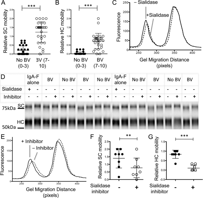
Electrophoretic mobility changes of SIgA caused by BV clinical samples can be prevented using the sialidase inhibitor dehydro-deoxy-sialic acid. SIgA-F was mixed with material eluted from vaginal swabs from women with or without BV (numbers in parentheses indicate Nugent scores) to give 50 mm sodium acetate, pH 5.5, and 0.1 mg/ml SIgA-F. The mixture was incubated for 16 h at 37 °C, then denatured, and loaded onto a denaturing polyacrylamide gel. **, p < 0.005; ***, p < 0.0001. A and B, exposure to BV samples frequently resulted in faster migration of SIgA SC and HC. The graphs are metadata from 20 independently scored BV-positive and 20 BV-negative samples. C, histogram of IgA fluorescence versus migration distance following incubation in a representative BV-positive and matched BV-negative specimen. D, representative gel showing controls and specimens with and without the addition of 500 μm sialidase inhibitor (DDSia). E, a representative histogram of SIgA fluorescence versus migration distance in the presence or absence of DDSia. F and G, processing of IgA secretory component (F) and heavy chain (G) in BV specimens is reduced by inhibitors. In contrast, parallel incubation of SIgA-F with normal vaginal samples yields no apparent change in mobility, and the addition of sialidase inhibitor had no effect.

